At a Glance
| Topic | Key Facts |
|---|---|
| Procedure type | Two-level lumbar spinal fusion (L4–L5 and L5–S1) |
| Common indications | DDD, spondylolisthesis, spinal stenosis, disc herniation |
| Surgical approaches | PLIF, TLIF, ALIF (LLIF not suitable at L5–S1) |
| Hospital stay | Typically 2–4 days |
| Return to desk work | 4–6 weeks post-op (surgeon-dependent) |
| Full bone fusion timeline | 6–18 months |
| Success rate range | 65–90%, depending on patient factors and approach |
| Pseudoarthrosis (failed fusion) risk | Up to 15% in primary lumbar fusions |
| Adjacent segment disease (10-year) | ~31% after two-level fusion |
| Smoking and fusion failure | Significantly higher non-union risk in active smokers |
If you have already been told you need L4–L5–S1 spinal fusion surgery, you probably have a lot of questions that go beyond what a brief office visit can answer. What does the hardware actually look like inside the spine? What is happening to your bones in the months after the OR? When can you drive, or travel, or lift your grandchildren again? This guide is built around those exact questions, written for patients who are past the "should I have surgery?" conversation and now need to understand what this experience actually looks like, week by week and year by year.

What Is L4–L5–S1 Spinal Fusion, and Why Is It Performed?
L4–L5–S1 spinal fusion is a two-level procedure that permanently joins three vertebrae, L4, L5, and S1, into a single, stable structure. Unlike a single-level fusion at only L4–L5 or only L5–S1, a two-level fusion addresses pathology at both of those disc spaces simultaneously, which is why it requires more hardware, more bone graft material, and generally a longer recovery.
The lumbosacral junction, the place where the lumbar spine meets the sacrum, carries more mechanical load than almost any other region in the body. Every time a person bends, lifts, or simply walks upright, the forces generated by the torso converge at L4, L5, and S1. That persistent stress is precisely why these levels are also the most common sites for degenerative spine disease.
Anatomy of the L4, L5, and S1 Region
The L4 vertebra sits two levels above the sacrum and is the origin point of several major nerve roots that run into the thigh and knee. L5 sits just above the sacrum and contributes to the sciatic nerve, making it a frequent culprit in shooting leg pain and foot weakness. S1 is the top segment of the sacrum and anchors the entire lumbar column; its nerve roots govern sensation along the outer calf and the heel. When any of these three segments are diseased, the consequences can extend well into the legs, making simple daily activities genuinely difficult.
The Four Conditions Most Likely to Require This Surgery
Degenerative disc disease (DDD) is the most common indication. Over time, the discs between vertebrae lose their water content, flatten, and lose their ability to absorb shock. When two adjacent disc levels degenerate simultaneously, two-level fusion becomes the logical surgical response.
Spondylolisthesis is a condition in which one vertebra slides forward over the one beneath it. When this slippage occurs at L4–L5 and causes significant instability or nerve compression, fusion is often indicated. According to Mayo Clinic, spinal fusion is reserved for cases where conservative measures have not provided adequate relief.
Lumbar spinal stenosis refers to narrowing of the spinal canal that pinches the nerves within it. When stenosis at both L4–L5 and L5–S1 causes neurogenic claudication, a condition where walking triggers deep leg pain and weakness, surgical decompression paired with fusion can restore function.
Disc herniation with nerve compression, particularly when bilateral leg symptoms or progressive foot weakness are present, may require fusion in addition to simple discectomy when the disc space is significantly destabilized.
When Surgery Is the Right Choice (and When It Isn't)
Surgery is generally considered only after at least 6 to 12 weeks of conservative treatment, including physical therapy, anti-inflammatory medications, and targeted injections, have not provided meaningful relief. Cleveland Clinic notes that spinal fusion is typically reserved for more severe conditions or those unresponsive to non-surgical options. Emergency indications, such as rapidly progressing weakness or loss of bladder and bowel control, may require faster surgical timing. A doctor can advise on individual cases where the right timeline is unclear.
Before Surgery: What Your Spine Looks Like on Imaging
Before any incision is made, your surgical team builds a precise picture of your spine using multiple imaging modalities. Understanding what is actually shown on those scans can help you participate more meaningfully in your care decisions.
Reading Your Pre-Op MRI: Disc Degeneration, Stenosis, and Slip
An MRI (magnetic resonance imaging) scan is the clearest window into the soft tissue structures of the spine. On a pre-operative MRI for L4–L5–S1 disease, a radiologist is typically noting several findings.
Disc height loss appears as a narrowing of the space between vertebral bodies. In a healthy spine, each disc is clearly visible as a bright, fluid-filled cushion on T2-weighted MRI images. In a degenerated disc, that brightness fades and the disc appears flat and dark, a finding often described as "desiccation" or "black disc disease."
Foraminal narrowing refers to the tightening of the bony openings through which nerve roots exit the spinal canal. When a disc collapses, those openings shrink, and the nerve root that passes through them becomes pinched. The patient typically experiences this as radiating pain, numbness, or weakness in a specific pattern down the leg.
Nerve compression is visible when the disc material or bone spur has actually pressed against the nerve root itself. This appears on MRI as a loss of the normal clear border between the nerve root and the surrounding disc or bone tissue.
Spondylolisthesis grade is measured on a scale of I (mild) to IV (severe), with Grade I representing less than 25% slippage and Grade IV representing more than 75%. A Grade II or higher slip, especially when symptomatic, is a common prompt for two-level fusion.
What X-Rays Reveal About Vertebral Alignment Before Fusion
While MRI excels at showing soft tissue, plain X-rays, including flexion-extension (bending) views taken while standing, reveal dynamic instability. If a vertebra shifts significantly as you bend forward compared to backward, that motion suggests the disc can no longer stabilize the segment. That instability is one of the clearest indications for fusion.
X-rays also allow surgeons to measure lumbar lordosis, the natural inward curve of the lower back, and pelvic parameters, numbers that help them plan hardware placement to restore proper alignment rather than just locking the spine in whatever position it happens to be in.
Why Imaging Alone Doesn't Decide if You Need Surgery
Many people with dramatic MRI findings have minimal symptoms. Conversely, some people with relatively modest imaging changes have severe functional impairment. Surgeons use imaging findings in combination with the clinical examination, symptom severity, and response to conservative treatment to determine whether surgery is appropriate. Imaging is a map, not a verdict.
Preparing for Surgery: The 6-Week Prehabilitation Window
The 6 weeks before surgery matter far more than most patients realize. What happens in that window has a measurable effect on what happens in the months that follow.
Prehabilitation Exercises That Improve Fusion Outcomes
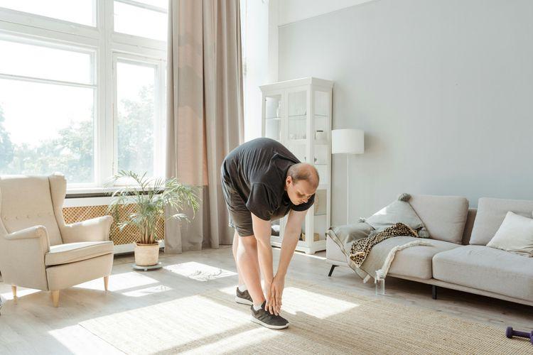
Prehabilitation, strengthening the muscles around the spine before surgery, is an approach with growing clinical support. The core muscles and hip flexors that stabilize the lumbar spine during recovery cannot be adequately trained while nerve pain is active post-operatively. Strengthening them ahead of time creates a reservoir of muscular support that carries the patient through the early recovery weeks when activity is most restricted.
A typical prehab program for lumbar fusion patients includes gentle core engagement exercises (dead bugs, pelvic tilts), walking programs to build cardiovascular base, and hip and glute strengthening to reduce the load transferred to the lumbar spine. A physical therapist familiar with spine surgery preparation can guide this process safely.
Medical Checklist: What to Do in the 6 Weeks Before Surgery
Per Cleveland Clinic pre-operative guidance, patients preparing for spinal fusion should expect the following in the weeks leading up to their procedure.
Lab work and medical clearance are standard and check for conditions such as uncontrolled blood pressure, clotting disorders, and kidney function, all of which affect surgical safety. A full medication review is required, with particular attention to NSAIDs (typically stopped 7 to 10 days before surgery due to bleeding risk), blood thinners, and any supplements that affect clotting. The surgical team will provide specific instructions about which medications to stop and when. Anesthesia planning, nutritional evaluation, and blood supply planning are also part of this phase.
Home preparation is worth treating seriously. Arrange a recovery area on the ground floor if possible, since stairs present a real challenge in the first week. Stock easy-to-prepare food, install a shower chair and grab bars, and raise toilet seat height if needed. Have a caregiver confirmed for the first 2 to 4 weeks.
Why Quitting Smoking Is the Single Most Important Pre-Op Step
Nicotine is a vasoconstrictor, meaning it narrows blood vessels and reduces the blood flow that bone tissue needs to heal and fuse. The research on smoking and spinal fusion failure is consistent and stark. A large meta-analysis found that the pseudoarthrosis rate, meaning failed bony union, was significantly higher in smokers than nonsmokers in lumbar fusion cases, with an odds ratio of approximately 1.97 in adjusted analyses, per a PMC-published systematic review. A separate study showed fusion failure rates as high as 40% in smokers undergoing primary spinal fusion surgery, according to data cited in ScienceDirect. Crucially, patients who stopped smoking at least one year before surgery showed fusion rates comparable to non-smokers. Quitting at any point before surgery reduces the risk; quitting well before surgery reduces it substantially.
Preparing Your Home for Recovery Before You Leave
Before the surgery date, set up a dedicated recovery station with everything within reach and at waist height to minimize bending and reaching. Include medication reminders, water, snacks, a phone charger, reading material, and any walking aid your surgical team recommends. The post-op instructions from your team will specify weight limits and restrictions; having the home configured before surgery means you will not have to improvise while in pain.
The Surgical Procedure: PLIF, TLIF, ALIF, and LLIF Compared
There is no single "right" approach to L4–L5–S1 fusion. The surgical technique your team selects depends on the anatomy of your specific pathology, your body type, prior surgical history, and the surgeon's training and preference. Understanding the major approaches helps clarify why your surgeon recommended one over another.
Approach Comparison Table: Posterior, Anterior, and Lateral
| Approach | Access Route | Best For | Key Notes |
|---|---|---|---|
| PLIF (Posterior Lumbar Interbody Fusion) | From the back | Most diagnoses; good nerve access | Longer muscle retraction; both-sided |
| TLIF (Transforaminal Lumbar Interbody Fusion) | From the back (one side) | Degenerative disc, spondylolisthesis | Less muscle disruption than PLIF |
| ALIF (Anterior Lumbar Interbody Fusion) | From the abdomen | L4–L5, L5–S1; large cage placement | Better disc space restoration; no back scar |
| LLIF (Lateral Lumbar Interbody Fusion) | From the side | Upper lumbar levels | Not used at L5–S1 due to pelvic anatomy |
The TLIF approach has become one of the most widely used for two-level lumbar fusions because it provides good access to both disc spaces while causing less disruption to the back muscles compared to traditional PLIF. The ALIF approach, accessing the spine through a small abdominal incision, allows surgeons to place a larger cage that can more fully restore disc height and lumbar lordosis, but requires coordination with a vascular or general surgeon for access. The LLIF approach cannot be used at the L5–S1 level because the iliac crest blocks lateral access at that location.
Bone Graft Options: Autograft, Allograft, and Biologics
Bone graft material is what actually produces the fusion. Without a biological scaffold for new bone to grow through, the vertebrae will not fuse regardless of how well the hardware is placed.
Autograft, taken from the patient's own body (often the iliac crest or locally from the surgical site), remains the gold standard for fusion because it provides both the structural scaffold and the live bone cells needed for growth. The tradeoff is a secondary donor site, which adds some recovery burden.
Allograft, cadaveric bone that has been processed and sterilized, eliminates the donor site but provides only the scaffold without living cells. It is frequently combined with the patient's own bone marrow aspirate or growth factors to improve biologic activity.
Synthetic biologics, including recombinant bone morphogenetic protein (rhBMP-2), have shown promise in improving fusion rates in specific populations, including smokers, according to research published via PMC.
What Robotic-Assisted Fusion Means for Precision and Outcomes
Robotic-assisted spine surgery uses pre-operative imaging to create a three-dimensional map of the spine, which the robot's guidance system then uses to direct pedicle screw placement with sub-millimeter precision. Early outcome data suggest robotic assistance reduces the rate of misplaced screws, which can cause nerve injury or require revision surgery. It is not universally available and does not change the fundamental biology of fusion; it improves the precision of hardware placement.
After Surgery: What Your Spine Looks Like on Imaging
The "after" side of the before-and-after picture is actually a slow-motion transformation that unfolds over 12 to 18 months. Understanding what each set of post-operative images is showing helps set realistic expectations at each checkup.

Post-Op X-Ray: What the Hardware Looks Like in Place
The first post-operative X-ray is typically taken before you leave the hospital. What you will see on this image may initially look alarming, but it represents the mechanical scaffolding that holds the fusion construct in place while bone grows.
Pedicle screws, cylindrical titanium screws inserted into the pedicles (the bony stalks) of each vertebra, are the anchor points. Connecting rods link the screws on each side, forming a rigid frame. Between the vertebral bodies sits the interbody cage, a titanium or PEEK (polyether ether ketone) spacer packed with bone graft material. This cage acts like a tent pole, holding the disc space open at a height more consistent with the pre-degeneration state and providing a chamber for bone graft to grow into a solid column.
On X-ray, the restored disc height is visible, particularly in ALIF cases where cage size allows for more aggressive height restoration.
6-Month MRI: Confirming Bone Fusion Has Occurred
At around 6 months post-operatively, if fusion appears to be progressing well clinically, surgeons may order a CT scan rather than MRI, because CT better visualizes new bone formation around and through the cage and hardware. On CT, solid fusion appears as a continuous bridge of bone linking the vertebral bodies. This bridging bone is what a "successful fusion" actually means.
On MRI at the same timepoint, reduced nerve compression is often visible as the nerve root now has space around it that was not present before surgery. Swelling and surgical changes near the fusion site are expected and gradually resolve over the first year.
The standard imaging checkpoints after L4–L5–S1 fusion are typically: X-ray at 6 weeks (hardware position check), X-ray or CT at 3 months (early fusion assessment), CT at 6 months (bone bridging confirmation), and X-ray at 12 months (long-term alignment review).
What a Failed Fusion Looks Like on Imaging (and Warning Signs)
Pseudoarthrosis, or failed bony fusion, is visible on CT as a persistent gap or lucent halo around the screws or cage rather than solid bone bridging. On X-ray, dynamic studies (flexion-extension views) may show abnormal motion at the fused levels, a sign the spine has not stabilized as intended.
Hardware failure, meaning a fractured pedicle screw or broken rod, appears on plain X-ray as a break in what should be a continuous metal structure. Hardware failure is often a sign of pseudoarthrosis because intact hardware rarely breaks in a solidly fused spine. The forces that cause hardware fatigue fracture are the same cyclical loads that should have been absorbed by mature bone.
Warning signs that a fusion may not be healing correctly include persistent or worsening back pain beyond 6 months, pain that increases with activity rather than improving, new or returning leg pain, and visible hardware changes on follow-up imaging. These findings warrant prompt consultation with the surgical team. If you notice any of these signs, connecting with a specialist through Momentary Lab's physician directory can help you find a qualified spine surgeon for a second opinion or evaluation.
Week-by-Week Recovery Timeline: Hospital to 12 Months

Recovery from a two-level lumbar fusion is not a straight line. There are phases of rapid improvement, frustrating plateaus, and gradual milestones that only become clear in hindsight. The timeline below is a clinical framework; individual progression varies depending on age, health status, and surgical approach.
Days 1–3: Hospital Stay and First Steps
Surgery typically lasts 3 to 6 hours for a two-level fusion, after which the patient wakes in a recovery unit and is usually moved to a standard room within a few hours. The first walk, with a physical therapist at the bedside, often occurs on the day of or the day after surgery. This early mobilization is deliberate: blood clots are a serious post-operative risk, and walking is one of the most effective preventive measures.
Pain in the immediate post-operative period is a combination of the old, familiar nerve pain (which may persist for weeks as the nerves heal) and new surgical pain from the muscle dissection. Many patients are surprised to find that the burning sciatica that had dominated their lives before surgery is significantly reduced or gone even in the hospital. The nerve, once decompressed, begins healing relatively quickly.
A urinary catheter is standard for the first day. Drain tubes at the surgical site may be present depending on the approach. Most patients are discharged on Day 2 or 3 with oral pain medications, a blood thinner to prevent clots, and written discharge instructions.
Weeks 1–6: The No-BLT Rule and Home Recovery
The first six weeks of home recovery are governed by a principle called the No-BLT rule: no Bending, no Lifting, and no Twisting. These three movements, forward flexion, axial loading, and rotational stress, are the exact forces that could disrupt the fusion construct before bone has had time to begin bridging. Strict adherence is not optional during this phase.
Walking remains the primary exercise during weeks 1 through 6. Begin with short walks (5 to 10 minutes) several times per day and gradually increase duration as tolerance allows. Wound care, pain management, and constipation prevention (a common issue with opioid pain medications and reduced activity) are the practical daily priorities.
A shower is usually permitted after wound staples or sutures are removed, typically at the 2-week mark, though the surgical team will give specific guidance. Stairs are generally allowed from the outset but should be taken slowly and with support. Sleeping on your back with a pillow under your knees, or on your side with a pillow between your knees, is recommended.
Weeks 7–12: Starting Physical Therapy
Formal physical therapy typically begins between 6 and 12 weeks post-operatively, once the surgeon confirms on X-ray that early fusion progress is appropriate. Physical therapy at this stage focuses on restoring range of motion, rebuilding core muscle endurance, and correcting movement patterns that may have developed as compensations for pre-surgery pain.
This is often the phase where patient frustration peaks. The acute surgical pain has resolved, but formal activity clearance is not yet granted. Patience here is directly rewarded: patients who complete a structured PT program rather than self-directing their return to activity consistently show better long-term outcomes.
Months 3–12: Bone Consolidation and Gradual Return to Life
By month 3, most patients with good early fusion progress are cleared for light activity expansion. Many return to desk or sedentary work around this time. Walking programs can extend to 30 to 60 minutes daily.
The bone graft material begins consolidating into solid bone over the 6 to 12 month window following surgery. This process, called osseointegration, is not visible to the patient but is measurable on imaging. By 6 months, CT scans in successful fusions typically show early bone bridging. By 12 months, the fusion mass is generally mature enough for clearance to more demanding physical activities.
Driving is typically approved around 2 to 4 weeks post-operatively for automatic vehicles (assuming the surgery was not on the right leg side), but this depends on whether the patient can perform emergency braking reliably and whether narcotics are still being taken.
Specific Activity Restrictions: A Practical Guide by Category
One of the most common frustrations after spinal fusion surgery is the absence of specific, organized guidance on when each activity can resume. The chart below reflects general clinical guidelines; always confirm timing with your surgical team, as individual circumstances vary.
Driving, Travel, and Everyday Activity Milestones
Driving is usually cleared at 2 to 4 weeks, assuming no narcotic pain medications are in use and the patient can perform emergency braking safely. Long drives should be broken up with stops for walking in the early months.
Air travel is generally discouraged for the first 6 to 8 weeks due to blood clot risk. After that window, aisle seating and regular in-flight walks are recommended.
Desk work is typically approved at 4 to 6 weeks, with gradual return to full-time hours. Ergonomic seating, lumbar support, and frequent standing breaks are strongly recommended.
Light housework, such as loading a dishwasher or folding laundry at a raised surface, is usually permitted around week 4 to 6. Vacuuming, which involves repetitive twisting, should be avoided for at least 3 months.
Sexual activity is a topic patients often hesitate to raise. General guidance places resumption at 4 to 6 weeks, but position selection (avoiding positions that require significant lumbar flexion or twisting) and surgeon confirmation are both important.
Permanent Weightlifting and Twisting Restrictions
L4–L5–S1 fusion creates a permanent alteration in lumbar mechanics. Many surgeons recommend a long-term lifting ceiling of 20 to 40 pounds, though this figure varies by the number of levels fused, patient age, bone quality, and the completeness of fusion. Repeated heavy overhead lifting and deep forward bending are biomechanically stressful on both the fusion construct and the adjacent segments above it.
Heavy lifting restrictions are not meant to prevent one-time incidents but to protect against cumulative stress on the fused construct and the L3–L4 level above it.
Sports and High-Impact Activities: What's Possible Long-Term
Low-impact activities including walking, swimming, cycling on a stationary bike, and yoga (modified) are generally cleared between 3 and 6 months post-operatively, subject to surgeon approval. Golf and recreational hiking are typically cleared at the 9 to 12 month mark.
High-impact sports involving running, jumping, or contact are generally not cleared until after 12 months and only after confirmed solid fusion on imaging. Contact sports, including competitive martial arts, football, and rugby, may carry permanent restrictions given the risk of hardware disruption or adjacent segment injury.
Adjacent Segment Disease: The Long-Term Risk You Need to Know
When two vertebral levels are fused, the levels above and below the fusion are asked to compensate for the mobility that has been removed. Over time, this increased mechanical demand can accelerate degeneration at those levels, a complication called adjacent segment disease (ASD).
What Is Adjacent Segment Disease and How Common Is It?
Adjacent segment disease is defined as the development of new, symptomatic pathology at the level directly above or below a fusion construct. The research on ASD after two-level lumbar fusion is sobering but important to understand in context.
A landmark study on posterior lumbar fusion outcomes reported that the overall annual incidence of ASD requiring further surgery was 2.5%, with 10-year prevalence rates of 16% after single-level fusion, 31% after two-level fusion, and 40% after three or four-level fusions, according to data published in PubMed. Patients younger than 45 had significantly lower ASD risk than those older than 60.
Stopping the fusion at L5 rather than extending it to S1 was associated with a 1.7-fold higher risk of symptomatic ASD compared to fusions that included S1, according to the same dataset. This finding is counterintuitive to many patients but reflects the biomechanical demands placed on the L5–S1 level when it is left mobile adjacent to a fused segment.
For patients having L4–L5–S1 fusion specifically, the adjacent level at risk is L3–L4, the disc and joint complex directly above the L4 screw.
Evidence-Based Ways to Reduce Your ASD Risk After Fusion
While ASD cannot be entirely prevented, its timeline and severity can be influenced by patient behavior after surgery.
Maintaining a healthy body weight reduces axial load on both the fused segment and the adjacent levels. Regular core strengthening through physical therapy or a supervised exercise program supports the musculature that shares load with the spine. Avoiding high-impact repetitive stress, particularly heavy axial loading like regular weightlifting above the surgeon's recommended ceiling, reduces the mechanical demand on L3–L4. Good posture and ergonomic work setups matter, because prolonged flexion posture concentrates stress at the L3–L4 junction. Calcium, Vitamin D, and adequate dietary protein support bone density at the adjacent level, reducing the risk of end-plate fracture or rapid degeneration.
Success Rates, Outcomes, and Realistic Expectations
The term "success" in spinal fusion carries a specific clinical meaning that does not always match what patients hope it means. Understanding that definition before surgery prevents a great deal of post-operative disappointment.
What Does "Successful" L4–L5–S1 Fusion Actually Mean?
A successful fusion means two things: the vertebrae have achieved solid bony union, and the patient has experienced meaningful improvement in function and pain compared to their pre-surgical baseline. It does not mean complete elimination of all pain. Residual low back aching is common even after solid fusion, particularly in patients with long-standing pre-operative pain sensitization. The goal of fusion is stabilization and decompression, not a return to the spine of a 25-year-old.
Overall success rates for L4–L5–S1 fusion range from 65% to over 90%, depending on patient age, smoking history, bone quality, underlying diagnosis, and surgical approach, per a review of clinical literature including data published via PMC.
Signs Your Fusion May Not Be Healing Correctly
Pseudoarthrosis, or failure of the bone graft to bridge the vertebral bodies into a solid union, occurs in up to 15% of primary lumbar fusions. The signs that fusion may not be progressing correctly include persistent or increasing back pain after 6 months, new onset leg pain that was absent earlier in the recovery, pain that worsens with sustained walking or standing and does not gradually improve over weeks, and visible hardware changes or fracture on follow-up X-ray.
These signs require prompt imaging, often a CT scan, and re-evaluation by the surgical team. If pseudoarthrosis is confirmed, revision options include bone stimulation devices, graft augmentation, or revision surgery.
10-Year Outcomes: What Life Looks Like After L4–L5–S1 Fusion
Most patients who achieve successful fusion report sustained improvement in leg symptoms and meaningful improvement in their ability to walk, sleep, and participate in daily activities. Long-term data show that approximately 7.5% of patients require revision surgery within 10 years due to hardware failure, pseudoarthrosis, or adjacent segment disease, per published review data. That number is meaningful but also means that roughly 92% of patients do not require revision within the first decade. Life after two-level fusion is not pain-free for everyone, but for many patients it represents a genuine and durable improvement over the condition that prompted surgery.
Mental Health, Nutrition, and Holistic Recovery Support
The physical timeline of spinal fusion recovery is well-documented. The emotional reality is less often discussed, which is a gap worth filling because mental health has a measurable effect on surgical outcomes.
Managing Anxiety and Frustration During a Long Recovery
A 6 to 12 month recovery is genuinely difficult, particularly for active people who define themselves by their physical capabilities. Anxiety about whether the fusion is healing correctly, frustration with the pace of progress, and situational depression during periods of restricted activity are all common. These reactions are not weakness; they are predictable responses to a significant physical event.
Connecting with others who have undergone similar procedures (online spine patient communities moderated by clinical staff) can provide perspective. Maintaining structured daily routines, even when activity is limited, supports psychological stability. If persistent low mood, sleep disruption, or anxiety are affecting daily function, discussing these symptoms with a primary care physician or mental health provider is appropriate and genuinely helpful. This aspect of recovery receives less attention than it deserves.
What to Eat (and Avoid) to Support Bone Fusion
Bone fusion is a biological process, and nutrition directly fuels it. Adequate calcium intake supports the mineralization of new bone. The NIH recommends 1,000 to 1,200 mg of calcium daily for adults, obtainable through dairy products, fortified foods, and leafy greens. Vitamin D is necessary for calcium absorption; deficiency is common and directly impairs bone healing. Protein provides the amino acids required for bone matrix formation, making adequate protein intake especially important in the first 3 to 6 months post-operatively.
Foods and substances to limit include alcohol (which impairs bone healing and interacts with pain medications), excessive caffeine (which can reduce calcium absorption at very high intakes), and heavily processed foods high in sodium, which increase urinary calcium loss. Anti-inflammatory foods such as fatty fish, berries, and olive oil may help manage post-operative inflammation, though the evidence for specific dietary protocols in spinal fusion patients remains an active area of research.
Sleep Positions and Ergonomic Adjustments After Surgery
Back sleeping with a pillow under the knees to reduce lumbar lordotic strain is the most recommended position in early recovery. Side sleeping with a pillow between the knees to keep the pelvis and spine aligned is the second-best option. Stomach sleeping places the lumbar spine in extension and should be avoided, particularly in the first 6 weeks.
For desk work, invest in a chair with genuine lumbar support, set the monitor at eye level, and use a sit-stand desk if possible. The goal is to avoid prolonged static postures in any position. Moving briefly every 30 to 45 minutes is beneficial regardless of which position is otherwise most comfortable.
FAQ
Is L4–L5–S1 fusion a major surgery?
Yes, L4–L5–S1 fusion is considered a major surgical procedure. It involves general anesthesia, multiple incision sites or one extended incision, placement of hardware across three vertebral levels, and a hospital stay of 2 to 4 days. Recovery extends over 6 to 12 months. That said, "major" does not mean "dangerous" in the hands of an experienced surgical team; it simply reflects the scope of the operation and the recovery commitment involved.
How long does L4–L5–S1 fusion surgery take to heal?
Hardware placement is complete on the day of surgery, but bone fusion is a biological process that takes 6 to 18 months. Most patients feel meaningfully better by 3 to 6 months, but CT confirmation of solid bone bridging typically comes between 6 and 12 months. Bone quality, smoking history, nutrition, and adherence to post-operative guidelines all affect how quickly and completely fusion occurs.
How will I feel 3 months after lumbar fusion?
At 3 months, most patients are off opioid pain medications and managing with over-the-counter anti-inflammatories as needed. Leg pain (sciatica or radiculopathy) is usually significantly improved or resolved by this point, assuming the nerve decompression was successful. Back pain and fatigue from the surgery itself are typically still present but declining. Many patients return to desk work around this time and are beginning formal physical therapy. The fusion is biologically active but not yet complete.
How successful is L4–L5–S1 fusion surgery?
Clinical success rates range from 65% to over 90%, with outcomes depending heavily on the underlying diagnosis, the patient's overall health, smoking status, and the quality of post-operative rehabilitation. Spondylolisthesis with nerve compression tends to show the most dramatic improvement after fusion. Patients with degenerative disc disease as the primary indication often experience meaningful but more gradual relief. A surgeon who is transparent about individual prognosis, rather than citing only best-case figures, is providing genuinely useful guidance.
What is the "No-BLT" rule after spinal fusion?
The No-BLT rule stands for no Bending, no Lifting, and no Twisting. These restrictions apply most strictly during the first 6 weeks after surgery, when the fusion construct is most vulnerable to disruption. Bending forward places excessive flexion stress on the hardware and graft; lifting before muscle strength is restored puts shear force on the construct; and twisting combines both rotational and flexion stresses that can dislodge the cage or stress the pedicle screws. Strict compliance with these restrictions during the early recovery phase is one of the most actionable things a patient can do to support a successful outcome.
Can I use the Momentary Lab tool to learn more about navigating spine care?
Yes. If you are researching your care options, finding the right specialist, or trying to understand your imaging reports, the Momentary Lab AI Healthcare Navigator can help guide you through complex health information in plain language.
References
- Cleveland Clinic: Spinal Fusion — Overview of spinal fusion indications, preparation, procedure, and recovery guidelines.
- Mayo Clinic: Spinal Fusion — Clinical overview of when spinal fusion is indicated and what the procedure involves.
- PMC (Glassman et al.): The Effect of Smoking on Spinal Fusion — Clinical evidence on smoking, fusion rates, and the role of rhBMP-2.
- ScienceDirect: The Effect of Smoking on the Fusion Rate of Spinal Fusion Surgery — Meta-analysis of fusion failure rates in smokers versus nonsmokers.
- PMC: Adverse Impact of Smoking on Spine Fusion and Patient-Reported Outcomes — Systematic review and meta-analysis on pseudoarthrosis risk in smokers.
- PubMed: Incidence and Prevalence of Surgery at Segments Adjacent to a Previous Posterior Lumbar Arthrodesis — ASD annual incidence of 2.5% and 10-year prevalence rates by number of levels fused.
- PMC: Failure in Lumbar Spinal Fusion and Current Management Modalities — Fusion success rates of 65–100% depending on patient and surgical factors; pseudoarthrosis diagnosis and management.
- PMC: A Comparative Radiographic Analysis of Fusion Rate between L4–5 and L5–S1 in a Single Level PLIF — Radiologic fusion rates for single-level PLIF ranging from 71% to 96%.
- NIH Office of Dietary Supplements: Calcium Health Professional Fact Sheet — Recommended daily calcium intake for bone health in adults.